
A baby is created by the union of an egg cell from a woman’s body and a sperm cell from a man’s body. This union is called fertilization and marks the beginning of pregnancy. Each egg and sperm cell contains half of the genetic material, or chromosomes, necessary for being human life. These chromosomes contain thousands of sections called genes, which determine the various characteristics of the child. The woman and man each contribute twenty three chromosomes to their child, making a total of forty-six. Because of the billions of possible combinations that can be produced by these forty six chromosomes and their thousands of genes, every child is unique.
The woman’s biological contribution begins in one of her two ovaries. Every month, an egg follicle ripens and swells in one of the ovaries. This ripening of the egg cell is initiated by a pituitary hormone. The walls of the follicle surrounding the ripening egg produce oestrogen, which causes the lining of the uterus, or womb, to thicken. The oestrogen also causes the cervical mucus to increase and to become more receptive to sperm. When the egg is mature, it bursts out of the follicle and is released near the fringed end of the fallopian tube. The release of an egg from an overy is called ovulation. Ovulation usually occurs 14 days before the next menstrual period, or about midway through the cycle.
The fallopian tubes are muscular canals lined with fine hairs, called cilia that move with a wave like action, drawing the egg into the tube and through it toward the uterus. At the same time that the egg is travelling through the fallopian tube, the follicle, stimulated by a pituitary hormone, begins producing progesterone, another hormone, which causes the uterine lining to chicken further. The progesterone also slows down contractions in the uterus, which facilitate implantation of the fertilized egg.
The biological contribution that the man makes to the baby begins with the production of sperm cells in his tests, which are two organs that hang outside his body in a sac of skin called the scrotum. Sperm cells, or spermatozoa, are produced in the semi niferous tubules with in the testes and are propelled into the epididymis for storage until ejaculation. As the spermatozoa pass from the epididymis through the vas deference to the urethra, secretions are added from the seminal vesicles, the prostate, and the Cowper’s glands. The purpose of these secretions is to provide a nourishing and fluid material that helps the spermatozoa move through the vagina, where they are deposited during intercourse.
The penis becomes erect during sexual excitement as blood pours into its layers of spongy material and the veins leaving the penis begin to constricts. With further excitement, the muscles around the seminal vesicles, the vas difference, and the prostate gland contract, driving the semen into the urethra. The muscles in the penis then contract and push the semen through the opening of the urethra. About 250 million to 500 million sperm ejaculated, although less than 500 will reach the egg.
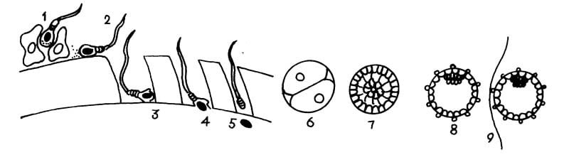
For fertilization to occur, sperm must reach the egg within 24 hours ovulation. Sperm remain viable for upto 72 hours. It takes sperm from 50 minutes to 2 hours to swim from the upper vagina, where they are usually deposited, to the outer third of the fallopian tube, where fertilization takes place. Sperm may reach an egg even if they are deposited externally, on the vulva. Only one sperm, out of the hundreds that may complete the journey, actually fertilizes the egg. As soon as this one sperm breaks through the egg wall, an enzyme releases that toughens the membrane of the egg and prevents penetration by any other sperm.
Implantation
The fertilized egg cell, now called an ovum, divides into two cells, then into four, and so on. By the fourth day, it is a cluster of sixteen cells called a morula. By the time it travels through the fallopian tube and reaches the uterus, a journey that takes 3 to 4 days, it is a hollow ball resembling a tiny blackberry and is known as a blastocyst.
About 8 days after fertilization, the blastocyst implants itself in the uterus, usually on the upper back wall. Projections on the outside of the blastocyst help it to attach to the thick inner lining of the uterine muscle. The blastocyst can then tap into the women’a blood supply for nourishment.
The Placenta
By the time the developing being is 2 weeks old, the placenta has begun to develop. The villi, embedded in the lining of the uterus, are forming primitive blood vessels, which tap into the woman’s blood supply. The function of the placenta is to supply the growing being with the oxygen and nutrients it needs from the woman’s blood system and to pass to the woman the waste products it does not need.
The fetal blood is always separate from the maternal blood. Substances are passed back and forth through a semipermeable membrane. At one time, the placenta was thought to act as a barrier to materials that might hurt the foetus. However, it is now known that almost everything that enters the woman’s body-including viruses, drugs, nicotine and alcohol-is passed to the foetus.
The placenta continues to grow until about 2 months before delivery, when it reaches its maximum size. At that time, it is about the size and shape of a dinner plate, approximately 8 to 9 inches in diameter, but thicker and heavier, weighing I to 2 pounds at birth. The side that is attached to the uterine wall is dark red and has sections like circular puzzle pieces in it. Scar like areas of tissue may be apparent. These are areas of calcification and they denote places that have degenerated. Women who smoke during pregnancy have more of these than do women who do not smoke. The side of the placenta that is next to the foetus is white and smooth, being covered by a membrane, the amniotic sac.
Nutrients coming from the woman pass to the blood vessels in the placenta. From the placenta, they move through the umbilical cord into the blood circulating within the foetus.
Inside the umbilical cord are three blood vessels-one large vein and two smaller arteries. Nutrients travel from the placenta to the foetus through the vein. Waste products return to the placenta through the arteries to be passed into the woman’s system. A jelly like substance called Wharton’s jelly surrounds the blood vessels and helps to protect them.
The umbilical cord begins to develop during the second week after conception and usually grows to about 18 inches in length, although it is possible for it to grow to anywhere from 12 inches to 40 inches. After the birth of the baby, the cord is clamped and cut. A stump remains, but dries up and falls off in 7 to 10 days.